Key Diagnostic Tests for Neuromuscular Diseases
By Jason Henninger | Friday, December 15, 2023
When a person visits their doctor with symptoms like weakness, muscle pain, or muscle cramps, the doctor might suspect a neuromuscular disease that affects muscles or the nerves that control them.
To help determine the cause of the symptoms and the best course of treatment, doctors often order diagnostic tests. Here are the tests most commonly used to diagnose neuromuscular diseases:
Creatine kinase (CK) and other blood tests
Blood tests may be used to check for certain enzymes, hormones, or other substances in the blood that could indicate a neuromuscular disease.
The CK measurement may be the most common blood test in the neuromuscular field. CK is an enzyme that is released into the bloodstream when muscles are damaged. Elevated CK levels could indicate muscular dystrophy or other muscle or nerve disorders. However, a high CK value does not confirm a diagnosis of a disease. It is usually followed with other diagnostic tests.
Blood tests may be used to look at levels of minerals and electrolytes, such as potassium, in the blood. High or low potassium can cause sudden attacks of muscle weakness or paralysis in periodic paralysis.
Antibody tests can be helpful in determining the presence of infection (as some neuromuscular diseases are viral in origin) or autoimmune diseases, such as dermatomyositis.
Electrodiagnostic tests
The electrodiagnostic test, which is often called EMG, has three main components:
- Nerve conduction studies that measure the electrical activity in nerves
- Electromyography (EMG) that tests muscle electrical activity at rest and during contraction
- Repetitive nerve stimulation to assess the function of the muscle-nerve junction
During nerve conduction studies, small electrodes are placed on the skin over specific muscles. These electrodes are connected to a machine that delivers small electrical pulses to the nerves and records their response. The electrical signals produced by the nerves can be measured, providing information about how well the nerves and muscles are functioning.
“EMG is a valid test to differentiate muscle diseases from diseases of the nervous system and junction between muscle and nerve,” says Margherita Milone, MD, PhD, a neurologist at the MDA Care Center at the Mayo Clinic in Rochester, Minnesota.
Genetic testing
Genetic testing is used to check for inherited neuromuscular diseases. This involves analyzing DNA samples from a cheek swab, blood sample, or tissue sample to look for specific gene mutations associated with certain disorders.
Genetic testing is considered the most accurate method for diagnosing a genetic disease. “If you have a genetic diagnosis [What’s the Difference Between a Clinical and Genetic Diagnosis?], that diagnosis could allow you to have a more specific treatment option than just treating overall symptoms. So, we do all the genetic testing that we can,” says Stephanie Acord, MD, a pediatric neurologist and director of neuromuscular disorders at Cook Children’s Hospital in Texas.
Lumbar puncture (spinal tap)
Lumbar puncture, also known as a spinal tap, involves inserting a thin needle into the lower back to collect some of the fluid that surrounds and protects the spinal cord. Information gathered from that fluid can help diagnose certain inflammatory conditions of the nervous system and autoimmune neurological conditions.
While lumbar puncture is generally considered safe, it can cause some discomfort. Local anesthesia is usually used to numb the area where the needle is inserted.
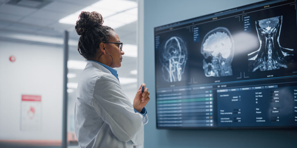
Magnetic resonance imaging (MRI) and computed tomography (CT)
MRI uses powerful magnets to capture images of the brain, blood, and bodily functions. CT uses X-rays to create 3D images of bones, tissues, and organs.
Both are safe, noninvasive imaging methods that can be used to diagnose a range of diseases. By providing detailed images of the internal structures of the body, they allow doctors to identify abnormalities or changes in muscles, bones, tissues, and organs. MRI and CT scans can identify specific areas of muscle or nerve damage, which can be critical for determining the type and severity of a neuromuscular disease.
In addition to aiding in diagnosis, MRI and CT scans can also be used to monitor the progression of a neuromuscular disease.
Muscle biopsy
This diagnostic procedure involves removing a small piece of muscle tissue and examining it under a microscope to look for abnormalities.
As noninvasive diagnostic techniques are improving, this procedure is being used less often to diagnose neuromuscular diseases.
“I would say muscle biopsy has really fallen out of favor,” says Jena Krueger, MD, a pediatric neurologist at the MDA Care Center at Helen DeVos Children’s Hospital in Michigan. “Most of us start with genetic testing because it’s sometimes as easy as a cheek swab.”
However, in some cases, this is still a useful diagnostic test. “A muscle biopsy remains crucial to make a precise diagnosis of most non-genetic muscle diseases or some mitochondrial muscle diseases,” says Dr. Milone.
Other tests
This list is not exhaustive — many other forms and versions of tests may be used to look for signs of a specific condition or rule out alternatives. For example, functional tests assess muscle strength, reflexes, coordination, and balance. Autonomic function tests look at part of the nervous system. Evoked potentials measure the brain’s responses to sensory stimuli and can give clues to nerve pathway issues.
There are numerous ways to look for a diagnosis, but the tests mentioned here are those one is most likely to encounter with a neuromuscular disease.
Next Steps and Useful Resources
- Learn more about understanding creatine kinase testing and levels.
- What’s the difference between a clinical and a genetic diagnosis? Find out here What’s the Difference Between a Clinical and Genetic Diagnosis?
- When you’re searching for a diagnosis, ask your doctor these questions.
- Stay up-to-date on Quest content! Subscribe to Quest Magazine and Newsletter.
Disclaimer: No content on this site should ever be used as a substitute for direct medical advice from your doctor or other qualified clinician.


